Medical Imaging for Early Cancer Detection
Head (AI Cloud Infrastructure), Presear Softwares PVT LTD
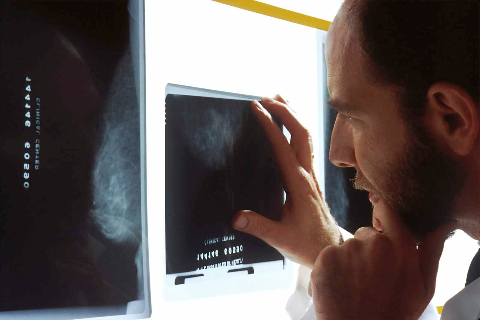
Introduction — the problem at hand
Early-stage tumors are often small, subtle, and easily missed in routine imaging. Radiologists work under heavy caseloads and variable image quality; manual reads can be inconsistent across facilities and shifts. Missed early detections mean later-stage diagnoses, more invasive treatments, poorer patient outcomes, and higher costs for hospitals and health systems. For radiology labs, oncology hospitals, and research institutes, the core pain point is clear: how to reliably detect cancer earlier while scaling throughput and maintaining—or improving—diagnostic accuracy.
Presear Softwares PVT LTD approaches this challenge by combining domain-focused machine learning, robust image processing pipelines, and clinician-centered workflows to create an end-to-end solution for medical imaging that aims to catch cancers earlier and consistently.
Solution overview
Presear’s Medical Imaging for Early Cancer Detection platform is a modular, secure system that augments radiologists’ workflows with algorithmic pre-screening, prioritization, and quantitative decision support. Rather than replacing clinicians, the system acts as an intelligent assistant that flags suspicious regions, scores risk, offers likely diagnoses, and integrates seamlessly into the existing radiology PACS (Picture Archiving and Communication System) and reporting workflows.
Key components:
Preprocessing & image harmonization: standardizes CT, MRI, and mammography images across scanners and protocols to reduce variability.
Detection & segmentation models: deep learning models trained on curated, annotated datasets to identify candidate lesions and precisely segment their boundaries.
Risk scoring & triage: interprets model outputs into clinically meaningful scores to prioritize cases that need urgent review.
Explainability & visualization: heatmaps, contour overlays, and measurement tools that help radiologists see why the model flagged a region.
Longitudinal tracking: tools for tracking lesions over time, detecting subtle growth trends that may indicate malignancy.
Integration & compliance: DICOM-compatible interfaces, role-based access, audit logs, and privacy-by-design to meet hospital and regulatory needs.
How it works — a practical workflow
Image ingestion: Scans (mammogram, chest CT, MRI, PET-CT) are routed to Presear’s preprocessing engine as soon as acquisition completes.
Normalization: The system applies noise reduction, intensity normalization, and geometric corrections so models receive consistent inputs.
Automated analysis: Detection models run and produce candidate regions with confidence scores and segmentation masks within seconds to minutes depending on modality and image size.
Triage & dashboarding: Cases are prioritized on a radiologist dashboard. High-risk cases move to the top of the reading list, with visual overlays and a concise summary of findings.
Radiologist review: The clinician reviews model output alongside raw images, accepts/edits segmentations, annotates, and finalizes the report.
Feedback loop: Edits and final reports feed back to Presear’s model-training pipeline to continuously improve performance in the specific institutional setting.
Longitudinal monitoring: Lesions are stored in a registry and tracked across timepoints with automatic comparison and growth metrics for care teams.
Technical notes — engineered for reliability
Model ensembles: Multiple architectures (e.g., encoder–decoder for segmentation, region proposal networks for detection) are ensembled to reduce false negatives.
Domain adaptation: Transfer learning with site-specific fine-tuning allows the system to adapt to the local scanner population and protocol mix.
Uncertainty quantification: Outputs include calibrated confidence measures. Low-confidence detections can be marked for double reads.
Edge and cloud deployment: Support for on-premises deployment to satisfy privacy or latency requirements, plus optional cloud-based model updates and analytics.
Interoperability: DICOM, HL7, and FHIR compatibility for integration with PACS, RIS, and EHR systems.
Benefits for stakeholders
For radiology labs
Increased sensitivity for early lesions: The system reduces the risk of missed small tumors by providing a second pair of algorithmic “eyes”.
Higher throughput: Automated pre-screening and triage reduce time-to-report for urgent cases and allow more efficient case distribution.
Quality assurance: Continuous performance monitoring, audit trails, and peer review facilitation improve QA programs.
For oncology hospitals
Earlier intervention: Tumors detected earlier are often more treatable with less aggressive therapy.
Resource optimization: Triage avoids delays for high-risk patients and enables better allocation of multidisciplinary tumor boards.
Data-driven care pathways: Quantitative lesion metrics support staging, treatment planning, and assessing response to therapy.
For research institutes
Cohort generation: Automated lesion detection accelerates the creation of research cohorts from imaging archives.
Reproducible annotations: Standardized segmentation and metadata make multicenter studies more reliable.
Clinical trials: The platform can help identify eligible patients earlier and track imaging biomarkers throughout trials.
Validation, safety, and compliance
Presear emphasizes rigorous validation:
Retrospective validation: Models are tested on held-out institutional datasets with metrics like sensitivity, specificity, AUC, Dice score for segmentation, and false negative rate analyses.
Prospective pilots: Before full deployment, Presear runs prospective validation in parallel with the standard-of-care workflow to measure real-world performance and workflow impact.
Human-in-the-loop: Radiologists retain final responsibility; the platform is explicitly designed to support decision-making, not to autonomously diagnose.
Privacy & security: Data encryption, access controls, and detailed audit logs ensure patient data confidentiality. Deployments can be configured to run fully on-premises to meet regional regulations.
Regulatory readiness: Presear prepares documentation and evidence packages to support institutional approvals and regulatory submissions as required by local authorities.
Implementation roadmap (practical steps)
Needs assessment: Presear conducts an on-site or virtual audit of imaging modalities, workflows, and IT landscape.
Pilot design: Define scope (modality, department), success metrics (e.g., sensitivity uplift, reporting time reduction), and governance.
Data curation & onboarding: Securely transfer de-identified historical data for model fine-tuning and validation; establish secure DICOM routing.
Integration & training: Integrate with PACS/RIS; train radiologists and staff on dashboards and feedback mechanisms.
Live pilot: Run the system in advisory mode (model suggestions visible but not enforced), collect metrics and user feedback.
Iterate & scale: Fine-tune models using local feedback, then expand deployment across departments and facilities.
Measurable outcomes (illustrative)
Institutions that adopt AI-augmented imaging can track outcomes such as:
Increase in early-stage detection rate (e.g., a measurable uplift in detection of small lesions on screening mammography or low-volume lung nodules on chest CT).
Reduction in diagnostic delay, measured as time from acquisition to finalized report for prioritized cases.
Reduction in unnecessary biopsies through improved specificity when segmentation and risk scoring support better clinical decisions.
Radiologist productivity gains, e.g., fewer minutes per case for cases where the model provides accurate pre-segmentation.
(Actual numbers vary by site and should be established during pilot evaluation.)
Real-world considerations & challenges
Data heterogeneity: Variations in scanners and protocols require careful normalization and ongoing monitoring.
Workflow adoption: Radiologist buy-in is essential. Presear emphasizes intuitive UI, transparent outputs, and measurable pilot results to drive adoption.
Regulatory approvals: Different countries and facilities have differing requirements; Presear supports customers through compliance processes.
Model drift: Continuous monitoring and periodic retraining help maintain performance as data distributions change.
Case study — hypothetical (illustrative)
A 600-bed oncology hospital integrates Presear’s platform for chest CT screening of high-risk patients. During a 6-month pilot:
The system triaged 12% of scans as high-risk; radiologists confirmed actionable findings in 70% of those.
Average time-to-diagnosis for prioritized cases dropped by 36%.
The hospital identified several sub-centimeter lung nodules that were stable on prior imaging but showed early growth trends; multidisciplinary review led to earlier biopsies and earlier treatment for a subset of patients.
This hypothetical demonstrates how early detection, improved workflows, and quantitative tracking combine to affect clinical care.
Conclusion — why Presear
Presear Softwares PVT LTD brings together machine learning expertise, practical engineering, and a clinician-first approach to address the critical need for earlier, more reliable cancer detection. By focusing on integration, explainability, and continuous improvement, Presear’s platform helps radiology labs, oncology hospitals, and research institutes convert imaging data into earlier diagnoses, better patient outcomes, and more efficient care delivery.
If your organization is evaluating AI for imaging, a focused pilot with clear success metrics—using Presear’s modular stack—can demonstrate value quickly while keeping clinicians in control. Early detection saves lives and costs; Presear’s approach is built to make that possible in real-world clinical settings.